A study at the University of Louisville James Graham Brown Cancer Center designed to make radiation treatment more accessible to women who face obstacles such as distance, transportation problems and time constraints is proving promising. The idea is to cut daily trips to the cancer center to once a week, says Dr. Anthony Dragon, a radiation oncologist at U of L. It also has the added benefit of cutting treatment costs by better than half.
Laura Ungar of The Courier-Journal in Louisville reports that Dragun led a previous study that found that "about a third of Kentucky women with early-stage breast cancer didn’t get recommended radiation treatments after lumpectomy surgery. Among those least likely to get radiation were rural Kentuckians, the elderly, African Americans and women in the Appalachian region of the state. Women who did not get recommended radiation were 60 percent more likely to die during the time they were studied."
Dragun told Ungar that the folks at U of L were not satisfied with just gathering those numbers, they wanted solutions to the problem. Dragun said the new experimental regimen is yielding good results, with women who get weekly radiation reporting similar levels of side effects as those getting radiation every day. Ungar notes that some outside experts say the approach does seems promising, but one critic was concerned about breast appearance results after the once-a-week treatments.
According the National Cancer Institute, more than 3,070 new cases of breast cancer were diagnosed in Kentucky in 2009 and was responsible for 614 deaths. (Read more)
Showing posts with label radiology. Show all posts
Showing posts with label radiology. Show all posts
Tuesday, November 27, 2012

Monday, June 20, 2011
Several Kentucky hospitals overdo CT scans, despite cost, risk
Despite the risk of exposing patients to more radiation and the fact that radiologists say it is very rarely necessary, Medicare outpatients are regularly receiving two CT scans on the same day at 12 Kentucky hospitals, according to analanysis of Medicare data by The New York Times, which produced a handy interactive map, a segment of which is copied here. To use the actual map, click on the map below.
![]()
Though only 5.4 percent of hospitals nationwide scan a patient's chest twice on the same day, nine Kentucky hospitals are double-scanning at least 15 percent of the time and three are doing it more than 30 percent of the time, accoding to data for 2008. Figures for 2009, which are expected to be similar, are due out next month from the federal Center for Medicare and Medicaid Services.
Though only 5.4 percent of hospitals nationwide scan a patient's chest twice on the same day, nine Kentucky hospitals are double-scanning at least 15 percent of the time and three are doing it more than 30 percent of the time, accoding to data for 2008. Figures for 2009, which are expected to be similar, are due out next month from the federal Center for Medicare and Medicaid Services.
Clark Regional Medical Center in Winchester has the highest rate, double scanning 43 percent of the time. Monroe County Medical Center in Tompkinsville double scans 34 percent of the time, followed by Pineville Community Hospital at 32 percent.
Other Kentucky hospitals that are double scanning at least 15 percent of the time include: Clinton County Hospital, Albany, 30 percent; Paul B. Hall Regional Medical Center, Paintsville, 25 percent; Harlan Appalachian Regional Healthcare Hospital, 24 percent; Norton Hospitals, Louisville, 23 percent; Westlake Regional Hospital, Columbia, 23 percent; Spring View Hospital, Lebanon, 20 percent; Methodist Hospital Union County, Morganfield, 19 percent; T.J. Samson Community Hospital, Glasgow, 18 percent; and The Medical Center at Bowling Green, 18 percent.
There are two types of CT scans, one that uses iodine contrast to monitor blood flow and one that does not. In rare circumstances, performing both scans — each with a separate charge — could help spot a tumor, said Dr. Michael J. Pentecost, a radiologist and Medicare consultant. While doctors may opt to double scan to get the most information on their patient possible, experts say being so thorough is rarely necessary, particularly given the added cost to the system and the exposure to radiation for the patient.
There are two types of CT scans, one that uses iodine contrast to monitor blood flow and one that does not. In rare circumstances, performing both scans — each with a separate charge — could help spot a tumor, said Dr. Michael J. Pentecost, a radiologist and Medicare consultant. While doctors may opt to double scan to get the most information on their patient possible, experts say being so thorough is rarely necessary, particularly given the added cost to the system and the exposure to radiation for the patient.
As Kentucky's numbers indicate, double scanning happens more often at small, community hospitals, where treatment can involve transferring patients to larger facilities. Nationwide, 200 hospitals administered double scans more than 30 percent of the time.
The Medicare agency released the data last year to hospitals "to show how they performed relative to each other and to encourage more efficient, safer practices," Walt Bogdanich and Jo Craven McGinty of the Times report. "The Medicare agency believes hospitals can and should do more to change physician behavior ... The federal agency plans to use other, similar measurements to rein in what it considers to be unjustified — and potentially dangerous — medical procedures." (Read more)
Thursday, March 17, 2011
Microsoft software to "replace" radiologists - recognizes organs and structures in medical images
Microsoft Research, Mar 8, 2011: InnerEye focuses on the analysis of patient scans using machine learning techniques for automatic detection and segmentation of healthy anatomy as well as anomalies:
Antonio Criminisi is the the researcher shown in the video above.
High-Performance Cancer Screening: See how a high--performance, 3-D rendering engine can be transformed into a real-world, life-saving medical application:
References:
Antonio Criminisi is the the researcher shown in the video above.
The InnerEye research project focuses on the automatic analysis of patients' scans by using machine learning techniques for:
- Automatic detection and segmentation of healthy anatomy, as well as anomalies
- Semantic navigation and visualization
Microsoft Research methods aim to combine medical expertise and modern machine learning theory in the design of tools for computer-aided diagnosis, personalized medicine, and natural user interfaces for surgical intervention.
The InnerEye project has a host of famous collaborators, including Johns Hopkins Medical Institute, The University of Oxford, Cornell Medical School, Massachusetts General Hospital, the University of Washington, Kings College London, and Cambridge University Hospitals.
High-Performance Cancer Screening: See how a high--performance, 3-D rendering engine can be transformed into a real-world, life-saving medical application:
References:
Comments from Twitter:
@hrana (Hisham Rana, MD): No thanks. RT @DrVes: Microsoft software to "replace" radiologists - recognizes organs and structures in medical images http://goo.gl/HwNNx
@doctorwhitecoat (Vamsi Balakrishnan): Definitely not a replacement... but cool tool in development.
@DrVes: Well, of course it's not a replacement... :) Just as "Watson" supercomputer won't replace PCPs/specialists...
@doctorwhitecoat (Vamsi Balakrishnan): Watson can't replace 3rd year Med students either; it can't do scutwork...:)
@ILoveOrthopedix (Orthopaedic Resident): MS Radiology! very interesting - the ECG machines recognise patterns & give diagnoses, but all the doctors make their own diagnosis.
@drcrosby (Bradley Dick): Reminiscent of Robin Cook's "Brain"?! (http://en.wikipedia.org/wiki/Brain_(novel))
Tuesday, March 30, 2010
GE Has Clinical IMAGES iPhone App for Radiologists
Clinical IMAGES from GE Healthcare showcases clinical images obtained from GE Healthcare imaging systems. You can choose a product and browse by anatomy or by applications specific to the selected system. The application contains both static images and video of various image acquisitions.
References:
GE Healthcare Releases Clinical IMAGES iPhone App for Radiologists
Clinical IMAGES for iPhone, iPod touch, and iPad
References:
GE Healthcare Releases Clinical IMAGES iPhone App for Radiologists
Clinical IMAGES for iPhone, iPod touch, and iPad
Sunday, March 14, 2010
Emphysema and Airflow Obstruction Associated with Impaired Left Ventricular Filling
 Very severe chronic obstructive pulmonary disease (COPD) causes cor pulmonale - elevated pulmonary vascular resistance and secondary reductions in left ventricular filling, stroke volume, and cardiac output.
The authors of this NEJM study hypothesized that emphysema, as detected on computed tomography (CT), and airflow obstruction are inversely related to left ventricular end-diastolic volume, stroke volume, and cardiac output among persons without very severe lung disease.
Of the study participants, 13% were current smokers, 38% were former smokers, and 49% had never smoked.
A 10% increase in emphysema on CT was related to decrease in left ventricular end-diastolic volume, stroke volume, and cardiac output.
Percent emphysema and airflow obstruction were not associated with the left ventricular ejection fraction (LVEF).

Mind map of Chronic Obstructive Pulmonary Disease (COPD) (click to enlarge the image).
References:
Percent Emphysema, Airflow Obstruction, and Impaired Left Ventricular Filling. R. Graham Barr et al. NEJM Volume 362:217-227 January 21, 2010 Number 3.
Image source: Enlarged view of lung tissue showing the difference between healthy lung and COPD, Wikipedia, public domain.

Wednesday, December 30, 2009
What is the radiation exposure from full-body scans used for airport security screening?
 There are 2 types of full-body scanner machines that use "weak" X-rays and radio waves respectively:
- Backscatter radiation X-ray full-body scanners. The image looks like a chalk drawing (shown right).
- "Millimeter wave" (radio wave) full-body scanners. The image looks like a fuzzy photo negative.
Backscatter radiation X-ray full-body scanners
The older type of full-body scanners use so-called backscatter radiation to scan the entire body to detect foreign objects. Passengers will be directed to stand against a refrigerator-size backscatter machine as a "pencil-thin" X-ray beam rapidly scans them to produce textured "charcoal outlines" of their bodies. The backscatter uses a narrow, low-intensity X-ray beam that scans the entire body at a high speed. The X-ray is not strong enough to penetrate much beyond the skin, so it cannot find weapons that may be hidden in body cavities.
The amount of radiation used during this scan is equal to 15 minutes of exposure to natural background radiation such as the sun's rays. One scan emits less than 10 microrem, the unit used to measure radiation. Comparably, an hour on an airplane at a high altitude exposes a passenger to 300 microrem, and the average person is exposed to 1,000 microrem of radiation over the course of a normal day.
Thirty hours of airplane travel is the equivalent of one chest X-ray (CXR) - an important health warning for frequent flyers.
A backscatter X-ray scan gives a person as much radiation as he or she would get from two minutes of flying in an airplane at 30,000 feet. A traveler would have to undergo more than a thousand scans in a year to equal one standard chest X-ray.
Dr. Albert J. Fornace Jr., an expert in molecular oncology at Georgetown University Medical Center, said such a low dose was inconsequential, even for pregnant women. “Obviously, no radiation is even better than even a very low level,” Dr. Fornace said. “But this is trivial.” But David J. Brenner, a professor of radiation oncology at Columbia University, said that even though the risk for any individual was extremely low, he would still avoid it.
"Millimeter wave" (radio wave) full-body scanners
The newer type of scanners, called a "millimeter wave" machine, doesn't use radiation. It uses electromagnetic waves to create an image based on energy reflected from the body. According to the TSA these devices deliver 10,000 times less energy than a person's cell phone.
The millimeter wave machine works like this: A person walks into a large portal that resembles a that resembles a glass elevator (9 feet tall and 6 feet wide), pauses and lifts his or her arms while the machine takes two scans using radio waves. The scans take 1.8 seconds, and it takes about a minute for the image to appear on a computer screen in a separate location.
Privacy concerns
The Transportation Security Administration (TSA) promises no one will see the revealing images except trained security agents staring at computer screens in a nearby room. The body scans will be deleted after 12 seconds.
Special “privacy” software intentionally blurs the image, creating an outline of a body that is clear enough to see a collarbone, bellybutton or weapon, but flattens details of revealing contours.
Lawyers for the American Civil Liberties Union (ACLU) have raised objections, calling the X-ray scan a “virtual strip-search.”
References:
Airport Scanner Radiation Risks Are Minimal, Government Report Says - NYTimes, 2012.
TSA to Conduct New Study on Safety of X-Ray Body Scanners http://goo.gl/r6Kfl
Dutch to use full body scans for U.S. flights. CNN.
New Airport X-Rays Scan Bodies, Not Just Bags. NYT.
For their eyes only? Boston Globe.
Manufacturer says full body scanners at airports are a valuable tool in fighting terror. The Plain Dealer.
Body-scan machine eyed for airports. AP.
Manchester airport trials naked-image security scans. Guardian.
Radiation risk low with whole-body airport scanners. Reuters, 2010.
New Airport Scanners: Radiation Risk Tiny. WebMD, 2010.
Image source: Wikipedia, backscatter X-ray, US Transportation Security Administration part of U.S. Department of Homeland Security, public domain.

Read more on a Kindle:
Sunday, November 8, 2009
BMJ video: The woman who knew too much
BMJ video: Alice Stewart: The woman who knew too much.
"Alice Stewart was one of Britain's foremost epidemiologists. However her recognition came late in her career, having spent her life fighting the establishment's enshrined views.
In the 1950s when she started her work, x-rays were routinely used in foetal monitoring. It was Stewart who first showed the link between the practice and childhood leukemia. She went on to look at the effects of low-level radiation exposure - uncovering the true adverse effects of chronic exposure, and thus earning herself the enmity of the nuclear industry."
Wednesday, September 30, 2009
Food Examples in Radiology
From Twitter account of @scanman, a radiologist in India:
 scanman Food examples in radiology - apple core, scalloping, berry aneurysm, lemon & banana (Chiari II), hamburger sign, coffee bean, dinner fork… scanman More… Onion skin (Ewing's), sausage digit (sarcoid), eggshell rice grain & popcorn calcification, pepper pot skull, honeycombing, donut… scanman My personal favourite is "licked candy stick appearance" of psoriatic arthropathy scanman Omental cake, pancake vertebra, celery stalk metaphysis, millet seed (miliary TB), cottage loaf sign (traumatic diaphragmatic rupture)
Related:
Selections from the Buffet of Food Signs in Radiology. Clare J. Roche et al. November 2002 RadioGraphics, 22, 1369-1384.
“Milk of Calcium” and Food Signs in Radiology. Lee A. Shratter, MD. May 2003 RadioGraphics, 23, 686.
Related:
Selections from the Buffet of Food Signs in Radiology. Clare J. Roche et al. November 2002 RadioGraphics, 22, 1369-1384.
“Milk of Calcium” and Food Signs in Radiology. Lee A. Shratter, MD. May 2003 RadioGraphics, 23, 686.
Saturday, September 12, 2009
Video: Routine Miracles with Dr. Conrad Fischer: Interventional Neuroradiology
"This book covers medical advances that would once have been called miracles but are now merely routine. The patients' stories within this book yield hope, optimism, and triumph. This is the best time to come out of medical school and training. This fact will inspire and uplift everyone in the medical profession as well as all of us who must, at some point, rely on the art of medicine to see us through." -- Conrad Fischer, M.D.
Visit http://seefisch.wordpress.com for more information about Dr. Fischer and Routine Miracles or follow Dr. Fischer on Twitter: @SeeFisch
See all videos of Routine Miracles with Dr. Conrad Fischer on YouTube: http://bit.ly/2TPUkU
Monday, February 16, 2009
Radiology Atlas for iPhone and iPod touch: Joslin Chest Atlas
I met Jeremy Joslin while attending the NEJM Horizons Conference in October 2008 and was impressed by his drive to change the way medical education is delivered. His idea of an ideal medical information tool was "a robot that sits on my shoulder all day while I am seeing patients and collects information. Then during my 30-minute commute home, I can ask him: "What would an expert do for this patient with new-onset Afib?," and it can make an MP3 of the discussion." Jeremy has not created this robot (yet) but his first step in this direction is impressive nevertheless:
The Joslin Chest Atlas is the first interactive Radiology Atlas for the iPhone and iPod touch

"By combining hundreds of clinician-years with iPhone magic, the Joslin Chest Atlas is able to provide a quite comprehensive atlas of common and uncommon radiographic findings of the chest.
Built with the medical student and resident in mind, this atlas demonstrates and quizzes you on must-know findings such as Hampton’s Hump, Kerley B lines, pneumomediastinum and more.
To make the magic sauce complete, we’ve expounded on each finding, providing a built-in link to Wikipedia and a hard-coded Google link to find out more.
Now that’s what we call osmotically-active software for your iPhone and iPod Touch.
Features Listing:
- 50+ radiographic findings in the chest
- Links to Wikipedia to read about each finding
- Tips for reading each finding
- High-resolution images with zoom in and out
- Quiz mode to test your reading abilities
- Pointers to identify features of each finding
- Send feedback, bug reports, or voice mail feedback from your iPhone
- Send iTunes App Store link to a friend from the app
- Frequent upgrades and new images
- Contribute your own images and get credit for your efforts
We need your help locating a radiograph that shows the Westermark sign! This rarely seen abnormality has resulted in a dearth of chest x-rays that are available (aka non-copyrighted) for us to include in the Joslin Chest Atlas. With your help, we can include this "phantom" sign into our program and thus, end the mystery that surrounds this finding. Think of this more like a bounty or treasure hunt, rather than an actual contest (some states and countries have weird rules about contests, and we don't feel like involving a team of lawyers). In exchange for your submission, we will reward you with $50.
Here are the rules:
- Send all submissions to contest@idiogenicosmoles.com
- Must be deemed as adequate quality. Generally, must be 640 pixels shortest side and not-overly compressed (uncompressed format is preferred if possible)
- Must be original film (not copyrighted) and not stolen off google images or such (we'll check!)
- Must be readily recognizable as Westermark Sign
- In case of multiple submissions, reward goes to first submitter with appropriate quality image. We reserve right to use any submitted film, however. In other words, if you send a poor image in before someone else sends in a great image, we may choose either at our discretion.
- Reward money paid via bank check.
- All decisions are final.
- Only 1 (one) bounty paid
- Deadline: April 1, 2009"
If you are interested in trying out and a writing a review for Joslin Chest Atlas, please contact me and I will email you one of the free codes Jeremy has provided me with.
The Joslin Chest Atlas is the first interactive Radiology Atlas for the iPhone and iPod touch

"By combining hundreds of clinician-years with iPhone magic, the Joslin Chest Atlas is able to provide a quite comprehensive atlas of common and uncommon radiographic findings of the chest.
Built with the medical student and resident in mind, this atlas demonstrates and quizzes you on must-know findings such as Hampton’s Hump, Kerley B lines, pneumomediastinum and more.
To make the magic sauce complete, we’ve expounded on each finding, providing a built-in link to Wikipedia and a hard-coded Google link to find out more.
Now that’s what we call osmotically-active software for your iPhone and iPod Touch.
Features Listing:
- 50+ radiographic findings in the chest
- Links to Wikipedia to read about each finding
- Tips for reading each finding
- High-resolution images with zoom in and out
- Quiz mode to test your reading abilities
- Pointers to identify features of each finding
- Send feedback, bug reports, or voice mail feedback from your iPhone
- Send iTunes App Store link to a friend from the app
- Frequent upgrades and new images
- Contribute your own images and get credit for your efforts
We need your help locating a radiograph that shows the Westermark sign! This rarely seen abnormality has resulted in a dearth of chest x-rays that are available (aka non-copyrighted) for us to include in the Joslin Chest Atlas. With your help, we can include this "phantom" sign into our program and thus, end the mystery that surrounds this finding. Think of this more like a bounty or treasure hunt, rather than an actual contest (some states and countries have weird rules about contests, and we don't feel like involving a team of lawyers). In exchange for your submission, we will reward you with $50.
Here are the rules:
- Send all submissions to contest@idiogenicosmoles.com
- Must be deemed as adequate quality. Generally, must be 640 pixels shortest side and not-overly compressed (uncompressed format is preferred if possible)
- Must be original film (not copyrighted) and not stolen off google images or such (we'll check!)
- Must be readily recognizable as Westermark Sign
- In case of multiple submissions, reward goes to first submitter with appropriate quality image. We reserve right to use any submitted film, however. In other words, if you send a poor image in before someone else sends in a great image, we may choose either at our discretion.
- Reward money paid via bank check.
- All decisions are final.
- Only 1 (one) bounty paid
- Deadline: April 1, 2009"
If you are interested in trying out and a writing a review for Joslin Chest Atlas, please contact me and I will email you one of the free codes Jeremy has provided me with.
Friday, January 9, 2009
CTisus.org: Free and Useful Radiology Education Website by Johns Hopkins
CTisus.com is created and maintained by The Advanced Medical Imaging Laboratory (AMIL) -- a team dedicated to research and education using medical imaging with a focus on spiral CT and 3D imaging.
The AMIL is part of the Department of Radiology at Johns Hopkins in Baltimore, MD.
A screenshot of the website is shown below:

The team behind CTisus.com also produces free vodcasts and podcasts: "Our lectures are now brought to you in Vodcast format. If you are already subscribed to the free CTisus Podcast, you will automatically receive our Vodcasts as well."
CTisus.com helps keep you up to date with the latest CT literature through the regular Journal Club and Imaging Pearls -- each month the editors provide summaries of key articles (12-20 per month) as well as a pearls section (25-75 per month) with bullet format key imaging information.
The AMIL is part of the Department of Radiology at Johns Hopkins in Baltimore, MD.
A screenshot of the website is shown below:

The team behind CTisus.com also produces free vodcasts and podcasts: "Our lectures are now brought to you in Vodcast format. If you are already subscribed to the free CTisus Podcast, you will automatically receive our Vodcasts as well."
CTisus.com helps keep you up to date with the latest CT literature through the regular Journal Club and Imaging Pearls -- each month the editors provide summaries of key articles (12-20 per month) as well as a pearls section (25-75 per month) with bullet format key imaging information.
Wednesday, March 12, 2008
The Times with an X-ray gallery of common ski injuries
 The winter is almost over but The Times of UK has just published an X-ray gallery of common ski injuries:
"If you are a skier, then the knee is the most likely injury on the slopes. And if you're a boarder? Do take care of your wrists, the most vulnerable to damage.
For skiers, the body parts most likely to be injured are the knee (32% of all ski injuries), head/face (13.7%), shoulder (9.1%), lower leg (7.6%), wrist (5.4%) and thumb (4.9%)"
References:
Ski breaks: the X-ray stories. The Times.
Image source: Bone fracture, from Wikipedia, the free encyclopedia (public domain).

Wednesday, January 30, 2008
Interesting Medical Websites
 I you are interested in using the web for medical education, you can always rely on DavidRothman.net and ScienceRoll to find something useful.
A few examples are listed below:
- GoldMiner is a radiology search engine. I added it to the list of resources on ClinicalCases.org: Imaging: Electrocardiograms, X-rays, CT scans. Link via DavidRothman.net.
- FreeMD is "an electronic doctor that conducts an interview, analyzes symptoms, and provides expert advice — for free.” The website may not be an "electronic doctor" but is simple and easy to use. In my tests, the online diagnostic tool FreeMD worked well, and due to its extensive use of pre-recorded videos, it felt more interactive. Link via DavidRothman.net.
Related:
Performance of a Web-Based Clinical Diagnosis Support System for Internists. Mark L. Graber and Ashlei Mathew. J Gen Intern Med. 2008 January; 23(Suppl 1): 37–40.
Image source: Wikipedia

Thursday, December 20, 2007
Free Online Atlases: Dermatology, Radiology and Ophthalmology
 Links:
Interactive Dermatology Atlas
Case-in-Point by American College of Radiology (ACR)
RedAtlas.org (Ophthalmology)
Ophthalmology Self-Guided Study Activity - ACP 2011 .
30,000 Medical Images, Tables and Figures from Leading Textbooks are Online. CasesBlog, 8/08/2005.
Free Ophthalmology Atlas. CasesBlog, 5/20/2007.
First 2 links are from MedSkool.
More Consequences of IT: The Disappearance of Radiology Rounds. Wachter's World, 01/2008.
30,000 Medical Images, Tables and Figures from Leading Textbooks are Online. CasesBlog, 8/08/2005.
Free Ophthalmology Atlas. CasesBlog, 5/20/2007.
First 2 links are from MedSkool.
More Consequences of IT: The Disappearance of Radiology Rounds. Wachter's World, 01/2008.
Friday, November 30, 2007
In the News: Serevent-related deaths and CT scans as a cause of cancer
 Some FDA panel members recommend Serevent be taken off the market
 Serevent, which is a part of the asthma blockbuster drug Advair, has been linked to sudden death in children. GSK claims there is no increased risk of hospitalization and mortality with Advair.
Serevent (salmeterol) is a long-acting beta2-adrenergic receptor agonist (LABA) which is used for treatment of asthma and COPD. The FDA will look into the safety profile of all LABAs at a future meeting.
Long acting beta-adrenoceptor agonists include salmeterol, formoterol (part of Symbicort) and bambuterol.
Note: The photo on the right shows a Seretide-labeled metered-dose inhaler (MDI). Seretide is the trademark for salmeterol in Europe.
CT scan may increase cancer risk

According to a NEJM review quoted in the NYTimes, in a few decades, 2 percent of cancers in the United States may be due to radiation from CT scans given now.
The average American’s total radiation exposure has doubled since 1980, largely because of CT scans. About 62 million of them were done in the U.S. in 2006 (up from three million in 1980) and studies suggest that a third of CT scans are unnecessary.
References:
Stronger Warnings Advised on Asthma Drug. NYTimes.
Report Links Increased Cancer Risk to CT Scans. NYTimes.
Current Concepts: Computed Tomography — An Increasing Source of Radiation Exposure. D. J. Brenner and E. J. Hall. NEJM.
Image source: Wikipedia, GNU Free Documentation License and public domain.


Thursday, June 28, 2007
What to do if you lose a guide wire during central line placement?
 I was discussing central line placement complications with medical students a few days ago. Some of them are illustrated with horrifying details in the NEJM.
"What should you do if you lose a guide wire during central line placement?", I asked.
"We could use a strong magnet to pull it out", one of the students suggested.
Well, not really.
You should call interventional radiology immediately and the radiologist will extract the wire under fluoroscopic guidance by using another wire to hook up and pull out the lost one.
A few examples of different approaches to extract lost guide wires are listed in the British Journal of Anaesthesia.
We have published free illustrated step-by-step procedure guides to central placement with and without ultrasound. The guides are available as web pages and Windows Mobile/Palm downloads.
References:
Central Line Placement: A Step-by-Step Procedure Guide with Photos
Loss of the guide wire: mishap or blunder? British Journal of Anaesthesia, 2002, Vol. 88, No. 1 144-146.
Lost guide wire during central venous cannulation and its surgical retrieval. Kumar S, Eapen S, Vaid VN, Bhagwat AR. Indian J Surg 2006;68:33-34.
Image source: Wikipedia, public domain.

Further reading:
Lost Guide Wire. Radiology Picture of the Day.
A Big Time Mistake. Ten out of Ten: My experiences as an ER doc, 02/2008.
Wednesday, January 10, 2007
Something you have never seen before -- a rare central line complication in NEJM
A man had a central line inserted by a cardiologist through the left subclavian vein. Six months later, the patient presented with posterior neck pain. A guide wire was protruding from the back of his neck... Continue reading: Complication of Central Venous Catheterization. NEJM, 2007.
Our own images of central line complications simply pale in comparison to this NEJM case.
You can get more cases and images if you subscribe to the free NEJM image feed which is syndicated in the right sidebar of this web site: http://rss.nejm.org/imagecast.xml
References:
Complication of Central Venous Catheterization. NEJM, 2007.
Complications of Central Line Placement - Pneumothorax, Arrhythmia, Hematoma

Our own images of central line complications simply pale in comparison to this NEJM case.
You can get more cases and images if you subscribe to the free NEJM image feed which is syndicated in the right sidebar of this web site: http://rss.nejm.org/imagecast.xml
References:
Complication of Central Venous Catheterization. NEJM, 2007.
Complications of Central Line Placement - Pneumothorax, Arrhythmia, Hematoma
Saturday, December 16, 2006
Radiology Picture of the Day
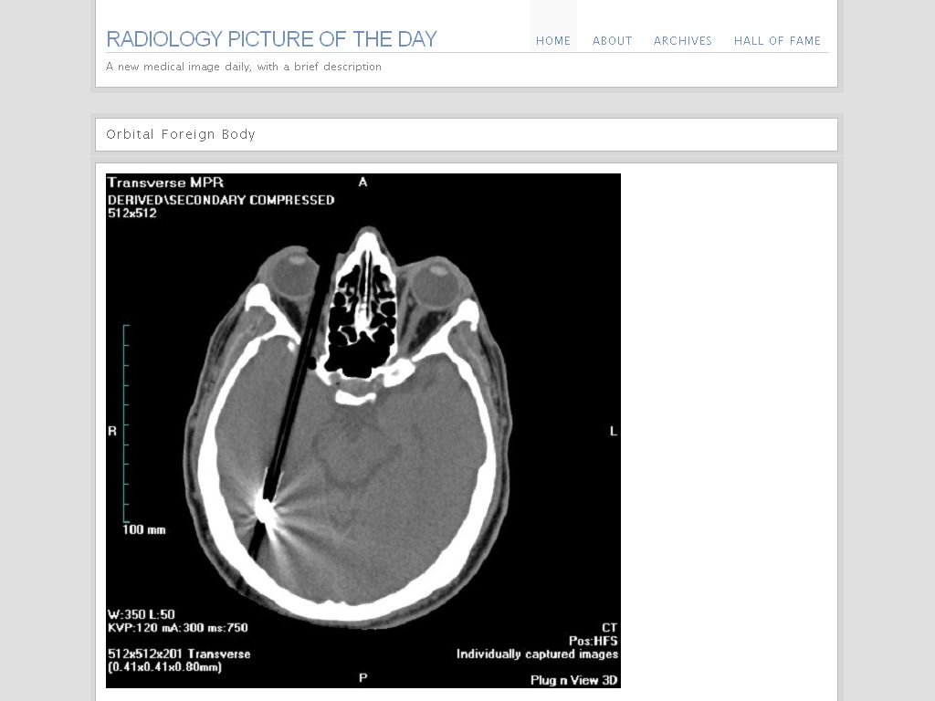 Radiology Picture of the Day features a new medical image daily, with a brief description. The website is edited and maintained by Dr. Laughlin Dawes who is a Radiology Registrar at Prince of Wales Hospital, Randwick. He lives in Sydney "with his wife Daniella, his sons Max and Alex, and Oscar the dog."
The website was started in 11/2006 and the Category Archives include:
Brain (10)
Cardiovascular (1)
Gastrointestinal (11)
Genitourinary (2)
Head & Neck (5)
Musculoskeletal (3)
Obstetrics (3)
Paediatrics (8)
Spine (2)
Thoracic (7)
There are more and more competitors to the famous NEJM Images in Clinical Medicine series...
References:
Link via Kevin, M.D.
NEJM RSS Feed for Image of the Week
Image source: Radiology Picture of the Day
Related:
More Consequences of IT: The Disappearance of Radiology Rounds. Wachter's World, 01/2008.
Updated: 01/31/2008
Subscribe to:
Posts (Atom)