
Showing posts with label Images. Show all posts
Showing posts with label Images. Show all posts
Saturday, April 23, 2011
The high cost of healthcare in America (infographic)
The high cost of healthcare in America (infographic) - click on the image to see the larger-size file. The site hosting the infographic, MedicalBillingandCoding.org, is one of the URL-grabbing portals that redirects to other sites, so be careful what you click there once you go beyond the image.

Thursday, March 17, 2011
Microsoft software to "replace" radiologists - recognizes organs and structures in medical images
Microsoft Research, Mar 8, 2011: InnerEye focuses on the analysis of patient scans using machine learning techniques for automatic detection and segmentation of healthy anatomy as well as anomalies:
Antonio Criminisi is the the researcher shown in the video above.
High-Performance Cancer Screening: See how a high--performance, 3-D rendering engine can be transformed into a real-world, life-saving medical application:
References:
Antonio Criminisi is the the researcher shown in the video above.
The InnerEye research project focuses on the automatic analysis of patients' scans by using machine learning techniques for:
- Automatic detection and segmentation of healthy anatomy, as well as anomalies
- Semantic navigation and visualization
Microsoft Research methods aim to combine medical expertise and modern machine learning theory in the design of tools for computer-aided diagnosis, personalized medicine, and natural user interfaces for surgical intervention.
The InnerEye project has a host of famous collaborators, including Johns Hopkins Medical Institute, The University of Oxford, Cornell Medical School, Massachusetts General Hospital, the University of Washington, Kings College London, and Cambridge University Hospitals.
High-Performance Cancer Screening: See how a high--performance, 3-D rendering engine can be transformed into a real-world, life-saving medical application:
References:
Comments from Twitter:
@hrana (Hisham Rana, MD): No thanks. RT @DrVes: Microsoft software to "replace" radiologists - recognizes organs and structures in medical images http://goo.gl/HwNNx
@doctorwhitecoat (Vamsi Balakrishnan): Definitely not a replacement... but cool tool in development.
@DrVes: Well, of course it's not a replacement... :) Just as "Watson" supercomputer won't replace PCPs/specialists...
@doctorwhitecoat (Vamsi Balakrishnan): Watson can't replace 3rd year Med students either; it can't do scutwork...:)
@ILoveOrthopedix (Orthopaedic Resident): MS Radiology! very interesting - the ECG machines recognise patterns & give diagnoses, but all the doctors make their own diagnosis.
@drcrosby (Bradley Dick): Reminiscent of Robin Cook's "Brain"?! (http://en.wikipedia.org/wiki/Brain_(novel))
Tuesday, October 19, 2010
Stereotypes in medical photographs
From BMJ:
If you search "medicine" on Google Images, you get a hundred million photographs.
The most common image is a stethoscope. The next is a bottle of pills (or, sometimes, red and black capsules). The next is a surgeon, masked and gowned, slicing skin with a scalpel.
Try "patient" and you will find lots of people in stripy pyjamas, lying obediently in bed, often with a spotty rash or a leg strung up in an orthopaedic hoist.
New images are needed to break the century-old stereotypes in medical photographs.
References:
New images needed: stereotypes in medical photographs. BMJ 2010;340:c1524.
If you search "medicine" on Google Images, you get a hundred million photographs.
The most common image is a stethoscope. The next is a bottle of pills (or, sometimes, red and black capsules). The next is a surgeon, masked and gowned, slicing skin with a scalpel.
Try "patient" and you will find lots of people in stripy pyjamas, lying obediently in bed, often with a spotty rash or a leg strung up in an orthopaedic hoist.
New images are needed to break the century-old stereotypes in medical photographs.
References:
New images needed: stereotypes in medical photographs. BMJ 2010;340:c1524.
Saturday, July 31, 2010
Not what it seems: Desert Island
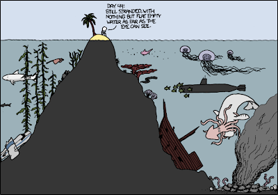
Not what it seems: Desert Island (click to enlarge the image). The obvious is sometimes hidden - this often applies to clinical evaluation of patients and research.
References:
http://xkcd.com/731
Thursday, July 8, 2010


Sunday, August 9, 2009
Impressive: SprintCam v3 showreel, made during a recent rugby competition in the Stade de France, Paris
I-Movix SprintCam v3 NAB 2009 showreel from David Coiffier on Vimeo. Go to Vimeo f0r the HD version of the clip.
Link via Dr. Shock who used to play rugby when he was younger.
Thursday, May 14, 2009
How to add NEJM Image Challenge gadget to your webpage
Here is how to add NEJM Image Challenge gadget to your webpage: http://bit.ly/e1KGL
There are more tools from the NEJM new projects list: http://bit.ly/RhqJi
Tuesday, April 14, 2009
Medical Q&A: What is shown in this picture?
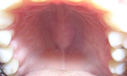
Medical Q&A: What is shown in this picture?
Highlight the white area below to see the answer:
Torus palatinus
This bony growth on the palate is the most common exostosis of the maxillofacial skeleton. It usually does not cause symptoms, but removal may be required if it interferes with the function or denture placement. Approximately 20% of the population has at least a small torus palatinus, which often goes unnoticed until middle age. This condition occurs twice as often in women as in men.
References:
MerckMedicus, SpringerLink, NEJM.
Image source: Wikipedia, Dozenist, GNU Free Documentation License.
Monday, January 19, 2009
Images in Medicine: Visualising Rhabdomyolysis
From The Lancet:
"A haemodialysis patient with hypercholesterolaemia was admitted with an ulcer on his left foot. Shortly after admission he complained of generalised aching and difficulty in mobilising.
Osteomyelitis complicating his foot ulcer was suspected and a Tc 99 -labelled diphosphonate bone scan was done. The scan showed uptake into damaged muscle"
Click here to see the images from The Lancet website.

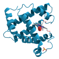
Model of helical domains in myoglobin, the protein linked to kidney damage in rhabdomyolysis. Image source: Wikipedia, public domain.
Rhabdomyolysis is the rapid breakdown (lysis) of skeletal muscle tissue (rhabdomyo) due to injury to muscle tissue. Rhabdomyolysis and its complications are major problems in people who are injured in disasters such as earthquakes and bombing. The disease and its mechanisms were first elucidated in the Blitz of London in 1941.
References:
Visualising rhabdomyolysis. Stephen Walsh et al. The Lancet, Volume 373, Issue 9658, Page 154, 10 January 2009.
Rhabdomyolysis, from Wikipedia, the free encyclopedia.
Related:
Rhabdomyolysis Review, Hospital Physician, 2009 (PDF).
Updated: 05/14/2009
"A haemodialysis patient with hypercholesterolaemia was admitted with an ulcer on his left foot. Shortly after admission he complained of generalised aching and difficulty in mobilising.
Osteomyelitis complicating his foot ulcer was suspected and a Tc 99 -labelled diphosphonate bone scan was done. The scan showed uptake into damaged muscle"
Click here to see the images from The Lancet website.
Model of helical domains in myoglobin, the protein linked to kidney damage in rhabdomyolysis. Image source: Wikipedia, public domain.
Rhabdomyolysis is the rapid breakdown (lysis) of skeletal muscle tissue (rhabdomyo) due to injury to muscle tissue. Rhabdomyolysis and its complications are major problems in people who are injured in disasters such as earthquakes and bombing. The disease and its mechanisms were first elucidated in the Blitz of London in 1941.
References:
Visualising rhabdomyolysis. Stephen Walsh et al. The Lancet, Volume 373, Issue 9658, Page 154, 10 January 2009.
Rhabdomyolysis, from Wikipedia, the free encyclopedia.
Related:
Rhabdomyolysis Review, Hospital Physician, 2009 (PDF).
Updated: 05/14/2009
Tuesday, December 2, 2008
CompFight is Beautiful Visual Search for Flickr
CompFight is a new visual search for Flickr which provides one of the best ways I have seen for browsing the site. It looks like most of the images are filtered by the tag "beautiful" or something similar because some of the photos are simply stunning.
The screenshot below shows the search results for "Cleveland":

The screenshot below shows the search results for "Cleveland":

Wednesday, November 19, 2008
LIFE photo archive hosted by Google includes millions of images, many historical medical photographs

The LIFE photo archive is hosted by Google which allows you to search millions of photographs stretching from the 1750s to today.
According to Google, "only a very small percentage of these images have ever been published. The rest have been sitting in dusty archives in the form of negatives, slides, glass plates, etchings, and prints. We're digitizing them so that everyone can easily experience these fascinating moments in time. Today about 20 percent of the collection is online; during the next few months, we will be adding the entire LIFE archive — about 10 million photos."
According to Google Blogoscoped, "as many photos are quite old, this also means many should have passed into the public domain zone, meaning you may be allowed to copy, edit, and republish that portion of the photos any way you like, including for commercial uses. Wikipedia says that “the copyright in a published work expires in all countries ... when ... The work was created and first published before January 1, 1923, or at least 95 years before January 1 of the current year, whichever is later.”
A few examples of image searches for medical terms are linked below - click to see the photos:
- heart attack, for example, Vice President Richard M. Nixon visiting Senator Lyndon B. Johnson at Bethesda Naval Hospital after Johnson's heart attack.
- cough
- bronchitis
- allergy
- diabetes
- headache
References:
LIFE Photo Archive available on Google Image Search. Google Blog.
Google’s Life Photo Archive. Google Blogoscoped.
Saturday, November 1, 2008
Last wave of summer on Flickr

The last wave of the summer, 2008

The last wave of the summer, 2007
Click here to see other great photos by Boby Dimitrov. All photos are under a Creative Commons license.
Thursday, August 21, 2008
Clinical Image: What is Bochdalek's Hernia?
In 1848, Bochdalek described the congenital defect in fusion of the posterolateral foramina of the diaphragm.

Bochdalek's hernia on the left

Bochdalek's hernia on the left
Most Bochdalek's hernias occur on the left side, allowing protrusion of the abdominal viscera into the chest. Previously unrecognized Bochdalek's hernia in adults is often diagnosed incidentally.
Read more here in Bochdalek's Hernia from Clinical Cases and Images.

Bochdalek's hernia on the left

Bochdalek's hernia on the left
Most Bochdalek's hernias occur on the left side, allowing protrusion of the abdominal viscera into the chest. Previously unrecognized Bochdalek's hernia in adults is often diagnosed incidentally.
Read more here in Bochdalek's Hernia from Clinical Cases and Images.
Wednesday, April 9, 2008
Wikis and Photo Sharing Explained in Plain English
Wikis in Plain English
Online Photo Sharing in Plain English
The authors from The Common Craft Show have published a series of short explanatory videos on different Web 2.0 (and other) topics. See more on YouTube.
Monday, March 31, 2008
Google Maps Street View Comes to Cleveland -- See Cleveland Clinic, UH, Metro, SVCH, etc.
Last week, Google Maps Street View expanded to more metropolitan areas in the U.S. and now includes Cleveland.
The Cleveland hospitals have become the "crown jewels" of the city. Some of the top health institutions in the country, and indeed, in the world, are located in Cleveland and now you can see them as if you there, from street level:
View Larger Map
This view from Euclid Avenue shows "the past and the future" of Cleveland Clinic. The original Clinic building is shown on the right and the new all-glass building of the Heart Institute is shown on the left.
View Larger Map
My office is in one of the buildings in the middle, called H. There are buildings from "A to Z" at Cleveland Clinic, and in fact, building A is shown below:
View Larger Map
The Crile building of Cleveland Clinic (building A)
View Larger Map
University Hospitals (UH)
View Larger Map
St. Vincent Hospital (SVCH)
Street View images like the ones above can easily be embedded in websites and blogs, so go ahead and share your favorites.
Video: How to use Street View of Google Maps
References:
Street View: expanding our horizons. Google Maps Blog.
Google puts its eye on Cleveland. The Plain Dealer.
Disclaimer: Dr. Dimov is a Clinical Assistant Professor of Medicine at Cleveland Clinic Lerner College of Medicine of Case Western Reserve University, Cleveland, Ohio, and a Staff Physician at the Department of Hospital Medicine, Cleveland Clinic, Cleveland, Ohio. All opinions expressed here are those of their authors and not of their employer.
The Cleveland hospitals have become the "crown jewels" of the city. Some of the top health institutions in the country, and indeed, in the world, are located in Cleveland and now you can see them as if you there, from street level:
View Larger Map
This view from Euclid Avenue shows "the past and the future" of Cleveland Clinic. The original Clinic building is shown on the right and the new all-glass building of the Heart Institute is shown on the left.
View Larger Map
My office is in one of the buildings in the middle, called H. There are buildings from "A to Z" at Cleveland Clinic, and in fact, building A is shown below:
View Larger Map
The Crile building of Cleveland Clinic (building A)
View Larger Map
University Hospitals (UH)
View Larger Map
St. Vincent Hospital (SVCH)
Street View images like the ones above can easily be embedded in websites and blogs, so go ahead and share your favorites.
Video: How to use Street View of Google Maps
References:
Street View: expanding our horizons. Google Maps Blog.
Google puts its eye on Cleveland. The Plain Dealer.
Disclaimer: Dr. Dimov is a Clinical Assistant Professor of Medicine at Cleveland Clinic Lerner College of Medicine of Case Western Reserve University, Cleveland, Ohio, and a Staff Physician at the Department of Hospital Medicine, Cleveland Clinic, Cleveland, Ohio. All opinions expressed here are those of their authors and not of their employer.
Saturday, January 12, 2008
Free Visual Dictionary of Medical Terms by Merriam-Webster
 The new Merriam-Webster's Visual Dictionary incorporates 6,000 color illustrations organized by subject area. The full-color pictorial images are grouped into chapters outlining major themes such as:
Earth
Plants & gardening
Animal kingdom
Human being
Food & kitchen
House
Clothing & articles
Arts & architecture
Communications
Transport & machinery
Energy
Science
Society
Sports & games
The free online access is heavily supported by ads but the medical section may still be useful to patients and students. The links include audio files with pronunciation.
References:
A Dictionary With a Point of View: Announcing Merriam-Webster's Visual Dictionary, Merriam-Webster, 2006.
Netter's Illustrated Human Pathology: A Searchable Collection of Images.
Image source: Merriam-Webster's Visual Dictionary
Updated: 01/22/2008
Thursday, December 20, 2007
Free Online Atlases: Dermatology, Radiology and Ophthalmology
 Links:
Interactive Dermatology Atlas
Case-in-Point by American College of Radiology (ACR)
RedAtlas.org (Ophthalmology)
Ophthalmology Self-Guided Study Activity - ACP 2011 .
30,000 Medical Images, Tables and Figures from Leading Textbooks are Online. CasesBlog, 8/08/2005.
Free Ophthalmology Atlas. CasesBlog, 5/20/2007.
First 2 links are from MedSkool.
More Consequences of IT: The Disappearance of Radiology Rounds. Wachter's World, 01/2008.
30,000 Medical Images, Tables and Figures from Leading Textbooks are Online. CasesBlog, 8/08/2005.
Free Ophthalmology Atlas. CasesBlog, 5/20/2007.
First 2 links are from MedSkool.
More Consequences of IT: The Disappearance of Radiology Rounds. Wachter's World, 01/2008.
Monday, December 17, 2007
NEJM Image Challenge and (Lack of) Wisdom of Crowds

The New England Journal of Medicine Image Challenge "provides an opportunity for you to use the clinical images from NEJM to test your diagnostic skills.
A new image and question appears each week, along with several possible answers. Before selecting an answer, you can take a closer look by zooming in and moving the image within the frame. You can also look at the relative distribution of answers chosen by other users." Source: NEJM.
The Image Challenge is a good review tool for USMLE, ABIM or residency in-service exams. When I studied for USMLE, I used to group clinical images from NEJM under system headings, e.g. Cardiology, Pulmonology, etc. but the image quiz/challenge is more fun.
The NEJM Image Challenge is, in a sense, a Web 2.0 tool -- it relies on user-created content in terms of recording readers' responses and comparing them to correct answers. It is surprising to see that for many image challenges the readers ("the crowd") did not get the right answer despite the massive participation of tens of thousands of responders for some queries -- click to enlarge the image above for an example. This may be an indication that for professional fields like medicine the "wisdom of crowds" concept does not work as well as, for example, Digg.com or Wikipedia. I would prefer an expert's opinion over the crowd's opinion most of the time, I think.
It could be an interesting mini-research project to have a look at all the answers in the NEJM Image Challenge and to see how many times the "crowd" got it wrong versus the "expert" (NEJM) who always provided the correct answer. In August 2007, I calculated the percentage of correct answers and the number of responses per question from all the images available on the NEJM website at the time. The results showed that there were on average 8300 responses per question and the "crowd" solved correctly only 69% of the image challenges.
Related reading
JAMA liked NEJM's Image Challenge so much that they launched their own "Clinical Challenge" http://jama.ama-assn.org/site/clinicalchallenge/jcg-index.xhtml
Make the Largest Encyclopedia in the World Better. I already did (I hope), 9/28/2005.
You know NEJM has a clinical "Image Challenge" right? Now The Lancet has "Picture Quiz" http://goo.gl/1NLIq - Where are you, BMJ?
Digg for Medicine: Nature Publishing Group Launches Dissect Medicine, 5/03/2006.
Edit Wars in Medical Wikipedia, 12/09/2006.
Google Finds Correct Diagnosis in 58 % of Cases Published in NEJM, 11/11/2006.
The only way to preserve the wisdom of the crowd is to protect the independence of the individual. WSJ, 2011.
NEJM Image Challenge is one of the most popular features on NEJM.org, and now there is a new way to play - on Facebook http://goo.gl/3VfGf
Digg for Medicine: Nature Publishing Group Launches Dissect Medicine, 5/03/2006.
Edit Wars in Medical Wikipedia, 12/09/2006.
Google Finds Correct Diagnosis in 58 % of Cases Published in NEJM, 11/11/2006.
The only way to preserve the wisdom of the crowd is to protect the independence of the individual. WSJ, 2011.
NEJM Image Challenge is one of the most popular features on NEJM.org, and now there is a new way to play - on Facebook http://goo.gl/3VfGf
Tuesday, November 27, 2007
Medicine 2.0 -- A Blog Carnival About Web 2.0 and Medicine
Medicine 2.0 is a weekly blog carnival which collects the most interesting posts about Web 2.0 and Medicine. The latest edition is posted here.


This week's host used blog screenshots and Slide.com very creatively -- see the embedded slideshow above. When I hosted, I tried something similar with Google Presentations but this looks much better.
Past editions and future hosts can be found on the blog carnival index page.
References:
Medicine 2.0, Blog Carnival About Web 2.0 and Medicine, Year 1, Issue 9
This week's host used blog screenshots and Slide.com very creatively -- see the embedded slideshow above. When I hosted, I tried something similar with Google Presentations but this looks much better.
Past editions and future hosts can be found on the blog carnival index page.
References:
Medicine 2.0, Blog Carnival About Web 2.0 and Medicine, Year 1, Issue 9
Friday, August 17, 2007


Subscribe to:
Posts (Atom)